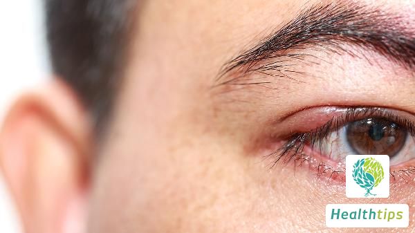
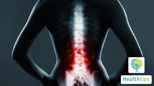
What Are the Causes of Bone Destruction?
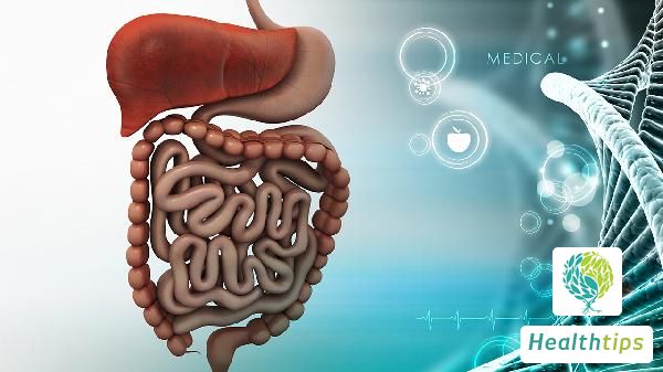
Bone destruction refers to the phenomenon of bone tissue loss in local bone caused by pathological reasons. Currently, there are four main types of bone destruction, namely subperiosteal type, osteolytic type, osteosclerotic type with limited osteolytic edge, and fragmentary type. There are many reasons for bone destruction, and everyone should be aware of the causes of this disease. So, what are the causes of bone destruction? Firstly, both bone cortex and cancellous bone can be destroyed. Secondly, it can be caused by inflammation, granuloma, tumor-like lesions, bone tuberculosis, skeletal trauma, pyogenic osteomyelitis, bone ischemia necrosis, trauma fractures, or residual bone in some malignant bone tumors. However, due to the nature of the lesion, the speed of its development, and the reactive changes of adjacent bone, the density of bone may be locally reduced, the trabecula may be sparse or bone defects may be formed, and the bone cortex may be destroyed, presenting a worm-eaten appearance. Secondly, what are the inspections for bone destruction? CT manifestations of osteolytic destruction include single or multiple bone dissolutions in the vertebral body, presenting as round or quasi-round low-density areas, some of which are cave-like or honeycomb-like, mostly without hardening edges, and with visible dead bone fragments inside. The typical manifestation is "nest-egg-like," which can break through the bone cortex to the surrounding area. CT manifestations of fragmentary destruction include collapse of the lesion vertebral body, loss of integrity into multiple bone fragments of varying sizes, shapes, and densities. The bone fragments may invade the epidural space and paravertebral tissues or be seen in the destruction areas of vertebral appendages and ribs. CT manifestations of subperiosteal destruction include worm-eaten or mouse-bite bone destruction at the anterior margin of the vertebral body, which can extend upward and downward along the subperiosteal and anterior longitudinal ligament to involve adjacent vertebral bodies. Paravertebral abscesses are often more evident. CT manifestations of osteosclerotic type with osteolytic edges include limited osteolytic destruction in the anterior half of the vertebral body with irregular morphology, obvious hardening edges, no dead bone found, and no obvious paravertebral abscess or only slight swelling of the paravertebral soft tissue.

Thirdly, how should bone destruction be prevented and treated? A rich diet should contain sufficient calcium, vitamin D, and other nutrients. Calcium and vitamin D are essential basic nutrients for increasing and maintaining bone mass. Protein and other nutrients such as phosphorus, sodium, and magnesium also play an important role in maintaining bone health. To increase calcium absorption, choose calcium-rich foods. For adults, the recommended daily calcium intake is 800 milligrams. Consciously add more milk and other dairy products to meals. You can also eat more seafood, shrimp, broccoli, green leafy vegetables, nuts, and dried fruits. Nutrients can also be obtained from pork, eggs, beans, and fish. Avoid excessive meat and salt as too much protein and salt can accelerate calcium loss. If you do not get enough calcium and vitamin D from your diet, you can take appropriate amounts of calcium supplements and vitamin D, especially for postmenopausal women. Among calcium supplements, calcium carbonate is the most commonly used, but it is not suitable for patients with hypochlorhydria. Organic calcium such as calcium citrate, although lower in calcium content, is more soluble than calcium carbonate and suitable for patients with hypochlorhydria. Calcium phosphate is not easily soluble and is not suitable for patients with chronic renal failure. People with renal dysfunction or who need to restrict certain nutrient intake should be more cautious when choosing.