Cervical Hypoechoic Lesion is a diagnosis report after a B-ultrasound examination. Through this examination, some abnormal manifestations in the cervical region can be detected, such as cervical hypoechoic lesions caused by cervical benign tumors, cervical cysts, and other factors. Therefore, when the inspection report indicates the presence of cervical hypoechoic lesions, attention should be paid, and further examinations should be conducted to screen for cervical cancer.
1. Cervical Hypoechoic Lesion
Cervical hypoechoic lesions are typically diagnosed after a B-ultrasound examination. A simple report of cervical hypoechoic lesions cannot determine whether it is cervical cancer. Cervical cancer needs to be confirmed through biopsy, colposcopy, and pathological examination based on the pathological results. Therefore, a simple cervical hypoechoic lesion may be cervical cancer, a benign tumor of the cervix, or a cervical cyst. In such cases, further examination is necessary.
2. Cervical Hypoechoic Lesion Examination
The issue of cervical hypoechoic lesions can be initially examined through a vaginal examination. After the cervix is opened with a speculum, the morphology of the cervix can be observed. If a more obvious lesion is seen visually, a biopsy can be taken directly. If the lesion is not very obvious, a cervical cancer screening should be performed, and the next step, whether a colposcopy is needed, will be determined based on the test results.
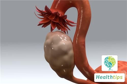
3. Low Echoic Lesion in the Cervical Canal
Low echoic lesions in the cervical canal may also be caused by submucosal fibroids that have prolapsed into the cervical canal or vagina. Low echoic lesions in the cervical canal are generally submucosal fibroids with long pedicles that have prolapsed into the cervical canal or vagina. The size, growth status, and length of the pedicle of the submucosal fibroid determine the differences in ultrasound and clinical manifestations.